

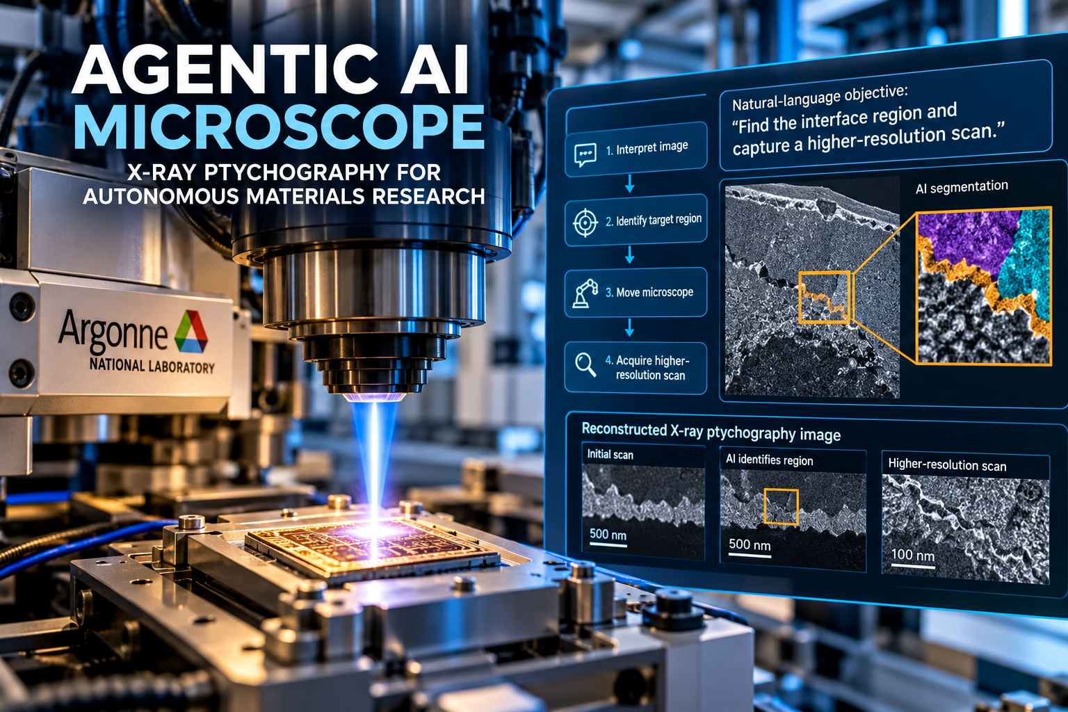
Agentic AI Microscope | Argonne National Laboratory researchers have demonstrated an agentic AI system able to guide parts of an X-ray microscopy experiment through natural-language instructions, shifting AI in microscopy from image analysis toward active experimental control.
The work comes from SYNAPS-I, short for Synergistic Neutron and Photon Science – Intelligence, a multi-laboratory project tied to the U.S. Department of Energy’s Genesis Mission. In a demonstration at the Advanced Photon Source, an AI agent received a high-level imaging objective, examined reconstructed data, searched for a target region inside a microelectronics sample, and directed the experiment toward a higher-resolution scan.
The important change is architectural. Traditional AI microscopy often analyzes data after acquisition. SYNAPS-I places an agentic layer inside the experimental loop, linking natural-language instructions, real-time image reconstruction, visual interpretation, instrument movement and follow-up acquisition.
This makes the Argonne system relevant to a broader shift toward autonomous microscopy and self-driving laboratories.
Quick Summary :-
Argonne’s SYNAPS-I demonstration combines three components:
• Real-time X-ray ptychographic reconstruction through PtychoFM.
• An agentic AI layer for experiment control and region-of-interest searches.
• Meta’s Segment Anything Model 3 for image segmentation.
The system tested a microelectronics sample at the 26-ID hard X-ray nanoprobe beamline. Low-risk actions run autonomously, while higher-risk or uncertain operations require human input. The demonstration is a research prototype, not a commercially available autonomous microscope.
What Is an Agentic AI Microscope?
An agentic AI microscope is a scientific imaging system in which AI performs more than image classification or post-processing. The AI receives a scientific objective, interprets observations, chooses among available actions, and sends instructions back into the experimental workflow.
This distinction matters.
A conventional AI-powered microscope might identify particles, segment structures, improve an image or classify defects. An autonomous microscopy system adds decision-making. It decides where to look next or which measurement to perform. An agentic system adds a broader reasoning layer, allowing a high-level objective to guide multiple steps in sequence.
Argonne’s approach follows this model. Researchers describe an imaging goal in natural language. The agent interprets reconstructed images, identifies relevant features and navigates toward a target region before initiating a more detailed scan.
A 2026 perspective in npj Computational Materials describes a similar direction for electron microscopy, with agentic systems supporting experimental design, closed-loop experimentation and real-time scientific reasoning.
What Did Argonne Demonstrate?
The demonstration took place at the 26-ID hard X-ray nanoprobe beamline at Argonne’s Advanced Photon Source. The facility supports nanoscale X-ray imaging, nano-diffraction and ptychography using hard X-rays.
The test focused on a microelectronics sample containing an interface between two regions.
Rather than providing a fixed sequence of microscope commands, researchers gave the AI a natural-language objective. The system started from a point inside the device and followed an iterative process:
Collect an image.
Interpret the reconstructed image.
Identify promising regions.
Move toward the target.
Refine the search.
Locate the region of interest.
Trigger a higher-resolution scan.
This process addresses one of the harder automation problems in microscopy: finding a feature whose appearance and position vary from sample to sample. Traditional scripts perform well when the workflow follows predictable rules. Region-of-interest searches often demand visual judgment and successive decisions.
How Does the Argonne AI Microscope Work?
SYNAPS-I connects several layers rather than relying on one AI model.
First comes real-time ptychographic reconstruction.
Ptychography records overlapping diffraction patterns and uses computation to reconstruct high-resolution images. At Argonne, the earlier SYNAPS-I phase used PtychoFM to reconstruct X-ray ptychographic images quickly enough to support experiment-time decisions.
Second comes the agentic control layer.
A multimodal large language model interprets reconstructed images and determines the next action within the allowed workflow. Instead of treating each image as the end of a pipeline, the system feeds visual observations back into the experiment.
Third comes image segmentation.
Argonne integrated Meta’s Segment Anything Model 3 into the workflow through the ALCF Inference Service. SAM 3 supports text and visual prompting for object detection, segmentation and tracking. Its role in SYNAPS-I adds another perception capability for isolating features inside reconstructed images.
The result resembles a closed feedback loop:
Natural-language objective → image acquisition → AI reconstruction → visual interpretation → decision → instrument action → new image.
This loop is the core technical significance of the demonstration.
Why Does Real-Time AI Matter for Microscopy?
Speed alone is not the main issue.
Modern synchrotron experiments generate enormous imaging datasets in short periods. Argonne reports millions of images from ultrabright X-ray experiments over a few hours. Earlier SYNAPS-I work focused on reconstructing ptychographic images during acquisition, reducing analysis delays and allowing researchers to alter measurements while an experiment remains active.
This changes the economics of experimental time.
Under a conventional workflow, researchers might collect data first, reconstruct it later, inspect the results and identify an important feature only after beam time ends.
An experiment-time AI system moves part of this decision process into acquisition.
For shared facilities, the effect could be significant. Beamline time is limited, instruments are expensive, and researchers often need to make decisions while measurements are running. Real-time reconstruction combined with adaptive control gives scientists more opportunities to redirect measurements before valuable experimental time expires.
DOE has placed similar goals inside the Genesis Mission, which targets AI-driven autonomous laboratories by combining robotics, edge AI, real-time analysis, intelligent feedback, hypothesis generation and scientific data management.
Is Argonne’s System a Fully Autonomous Microscope?
No.
The distinction is important for accurate reporting.
Argonne describes autonomous operation for low-risk actions, with human approval or guidance for higher-risk actions or situations involving greater uncertainty. The system therefore fits a supervised autonomy model rather than unrestricted machine control.
The announcement also presents the work as a demonstration and foundation for future self-driving microscopes, closed-loop experiments and automated defect detection. It does not present a commercial product, public deployment program, pricing model or general availability schedule.
This distinction should remain clear in coverage. Calling the system a fully independent research scientist would overstate the current evidence.
How Does This Compare With Other Autonomous Microscopy Work?
Argonne is entering an active research field rather than operating in isolation.
AILA, an Artificially Intelligent Lab Assistant, has been demonstrated on atomic force microscopy for tasks spanning experiment design, calibration, imaging and analysis. Its creators also introduced AFMBench to evaluate autonomous laboratory tasks and highlighted important limitations in current language-model agents, including instruction-following and safety concerns.
A separate 2026 npj Computational Materials perspective proposed “thinking microscopes” for electron microscopy, with specialized AI agents supporting experimental design, iterative closed-loop experimentation and real-time hypothesis generation.
Other systems target narrower use cases. SimuScan, for example, focuses on autonomous atomic force microscopy through synthetic-data-driven feature identification, segmentation and targeted imaging.
ALLocate takes another path by turning conventional microscopes into self-driving systems for acute leukemia detection, showing how autonomous imaging also has a diagnostic application.
The main differentiator in SYNAPS-I is its combination of natural-language control, real-time X-ray ptychography, multimodal agentic decision-making and segmentation inside a synchrotron workflow.
What Does Agentic AI Mean for Materials Science?
Materials science is a strong candidate for autonomous experimentation because researchers often work through repeated cycles of measurement, interpretation and targeted follow-up.
A material might contain a defect, interface, phase boundary or nanoscale structure worth investigating. Finding it through a large sample requires repeated measurements and expert decisions.
Agentic AI gives the experiment a mechanism for turning a scientific objective into a sequence of observations and actions.
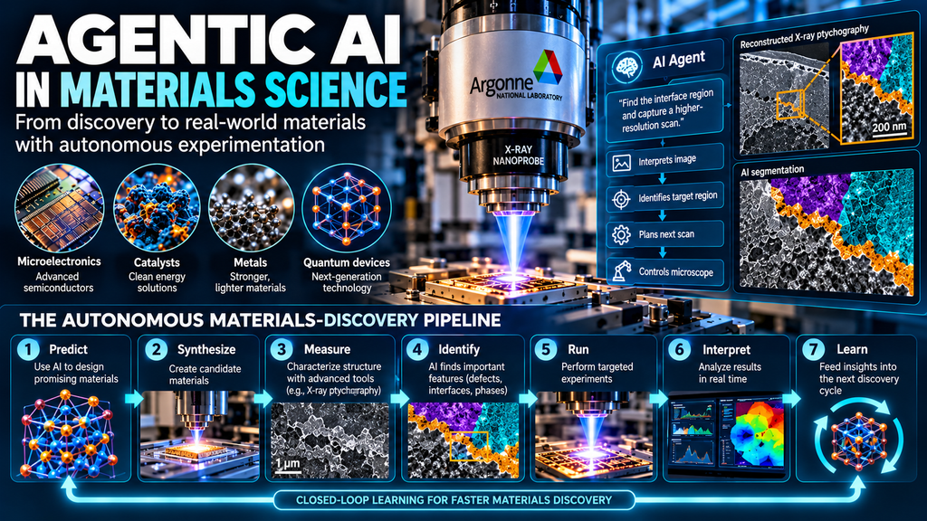
Argonne says the SYNAPS-I framework applies beyond microelectronics to catalysts, metals and quantum devices.
DOE’s broader materials strategy points in the same direction. Its Genesis Mission work describes systems linking prediction, synthesis, characterization and analysis into closed-loop learning systems for materials design and qualification.
This creates a broader research pipeline:
Predict a material.
Synthesize it.
Measure its structure.
Identify an important feature.
Run a targeted experiment.
Interpret the result.
Feed the result into the next research decision.
The microscope becomes one component within an autonomous materials-discovery stack.
What Are the Biggest Technical Challenges?
The hardest problems now sit beyond image recognition.
Instrument integration is one.
Scientific instruments need reliable interfaces for software control. The 2026 thinking-microscopes research argues for secure APIs, standardized metadata and stronger connections between instruments, computing systems and scientific data.
Data quality is another.
Agentic systems need access to high-quality experimental data, metadata, failed experiments and successful experiments. Without sufficient context, an AI agent risks making decisions based on incomplete information.
Safety is equally important.
A microscope operates physical hardware. Incorrect commands might waste beam time, damage a sample or move an instrument into an unsafe state. Argonne’s human-approval model addresses part of this problem, while prior autonomous-microscopy research shows why systematic benchmarking remains important.
Reproducibility also matters.
A useful autonomous laboratory system needs an auditable record of the objective, observations, decisions, actions and resulting data. Otherwise, faster experimentation could create a harder-to-review scientific record.
What Happens Next for Autonomous Microscopy?
The next step is likely broader closed-loop experimentation rather than simple microscope automation.
Argonne explicitly positions SYNAPS-I as groundwork for self-driving microscopes, automated defect detection and adaptive experimentation across scientific facilities.
The wider research community is pursuing similar ideas from different directions. Georgia Tech researchers describe agentic systems linked to electron microscopes for dynamic data collection and experiment prioritization.
The practical test will be repeatability.
A compelling autonomous microscopy system needs more than a successful demonstration. Researchers will need benchmark results across samples, experiments and laboratories, along with clear safety boundaries, failure reporting, reproducible workflows and instrument interoperability.
The strongest long-term model is therefore human-supervised autonomy.
Scientists define the research objective, constraints and acceptable actions. AI handles repetitive observation and decision cycles. Humans remain responsible for scientific interpretation, validation and high-stakes decisions.
For materials research, this division of work could shorten the time between an interesting observation and the next experiment.
FAQ About Agentic AI Microscopes
What is an agentic AI microscope?
It is a microscope or microscopy workflow in which an AI agent interprets observations, makes decisions and directs subsequent experimental actions toward a scientific objective.
What did Argonne National Laboratory demonstrate?
Argonne demonstrated SYNAPS-I at the 26-ID hard X-ray nanoprobe beamline, using natural-language instructions, real-time ptychographic reconstruction, agentic AI control and SAM 3 segmentation to locate a target region in a microelectronics sample and trigger higher-resolution imaging.
Is the Argonne microscope available commercially?
The accessible Argonne announcement describes a research demonstration. It does not provide commercial pricing, a product release date or general availability information.
What is ptychography?
Ptychography is an X-ray imaging technique based on overlapping diffraction measurements followed by computational image reconstruction. It supports high-resolution nanoscale characterization and is used at Argonne’s Hard X-ray Nanoprobe.
What is SAM 3?
SAM 3 is Meta’s image and video segmentation model, designed to detect, segment and track objects using text, exemplar and visual prompts. Argonne integrated it into the SYNAPS-I workflow for feature segmentation.
Will AI replace microscopy researchers?
The evidence supports a different direction. Current systems automate selected workflows while retaining human involvement for scientific judgment and higher-risk operations. DOE also describes Genesis as AI supporting researchers rather than replacing them.























Leave a Reply